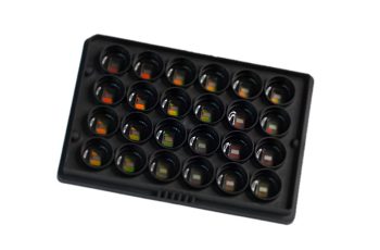
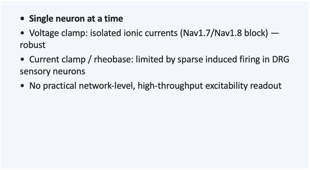
Dorsal root ganglion (DRG) sensory neurons rarely fire spontaneously, and evoked current-clamp rheobase protocols are constrained in this cell type, which has historically confined in vitro DRG pain pharmacology to voltage-clamp recordings of individual ionic currents: informative about target engagement, but silent on whether network-level excitability actually changes.
That gap matters because it shapes what a program can defend in a go/no-go decision. A Nav1.8 blocker that shows clean voltage-clamp current inhibition still leaves open the functional question: does it change how the network actually fires?
High-density microelectrode arrays (HD-MEA) are built to answer exactly that question, at a throughput patch clamp cannot match.
In practice, this has confined in vitro DRG pain electrophysiology to voltage-clamp recordings of ionic currents (e.g., Nav1.7/Nav1.8 current block) — informative on target engagement, but silent on the downstream question that matters most to a pain program: does the compound actually reduce the neuron’s capacity to fire?
HD-MEA (high-density microelectrode array) recording changes this picture.
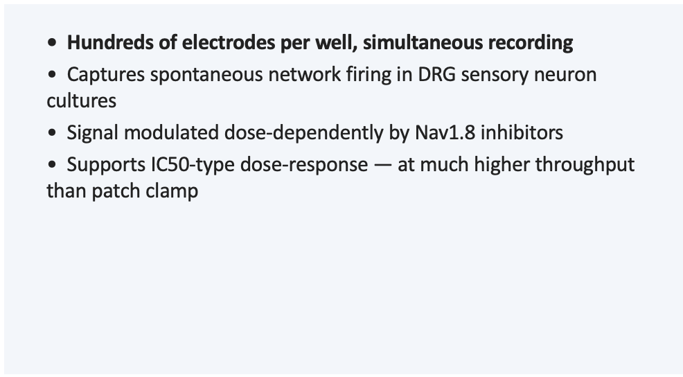
High density of electrodes per well + high electrode sensitivity lets us detect spontaneous network-level firing activity directly from cultured DRG sensory neurons — a signal that single-cell current-clamp protocols cannot reliably capture in this cell type.
• That signal is strong enough to be modulated pharmacologically by Nav channel blockers, including Nav1.8-selective inhibitors — enabling a dose-response, IC50-type readout at the network level.
Experiment 1: effect of a Nav1.8-selective inhibitor on DRG HD-MEA network activity
DRG cultures on HD-MEA plates develop stable baseline spontaneous network activity within 2–4 days in vitro (~100 active electrodes/well, firing rate stabilizing at 1–2 Hz).
• 500 nM TTX (pan-Nav blocker, positive control) did not significantly change firing rate at the group level despite the near-complete loss of active electrodes, consistent with a measurement-floor effect once very few electrodes remain active, not an absence of effect
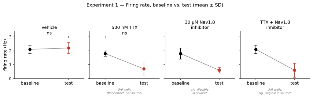
Firing rate (Hz), baseline vs. test — HD-MEA DRG culture, Experiment 1
Experiment 2: concentration-response of TTX and a clinically approved Nav1.8-selective inhibitor (suzetrigine)
After baseline characterization (stable network activity by 1–4 days in vitro), cultures were exposed to a concentration range of TTX or suzetrigine, a clinically approved Nav1.8-selective inhibitor.
• Both compounds produced clean, fittable concentration-response curves on the HD-MEA network readout (active electrode count, firing rate), with IC50 values in the expected pharmacological range for each tool.
• This is the key translational result: a network-level HD-MEA endpoint can resolve a dose-response relationship for a Nav1.8-selective, clinically validated compound, the core requirement for compound profiling in a pain discovery program.

Left: TTX concentration-response. Right: suzetrigine (Nav1.8 inhibitor) concentration-response. Source: NS USA internal HD-MEA DRG dataset (Sept. 2026).
Throughput, at an accessible cost point. HD-MEA records from large numbers of neurons in parallel, across a full 24-well plate, enabling a dose-response design (1 compound × 3 concentrations, ~80 compounds/month) or a compound-screening design (3 compounds × 1 concentration, ~240/month) — a markedly higher throughput than single-cell patch clamp.
The question facing most pain-focused discovery programs isn’t whether in vitro DRG electrophysiology belongs in the workflow, it’s whether the chosen readout can actually capture a functional, dose-dependent effect in a cell type that resists two of the field’s standard endpoints. Dorsal root ganglion (DRG) sensory neurons rarely fire spontaneously, and evoked current-clamp rheobase protocols are constrained in this cell type, which has historically confined in vitro DRG pain pharmacology to voltage-clamp recordings of individual ionic currents: informative about target engagement, but silent on whether network-level excitability actually changes.
That gap matters because it shapes what a program can defend in a go/no-go decision. A Nav1.8 blocker that shows clean voltage-clamp current inhibition still leaves open the functional question: does it change how the network actually fires? High-density microelectrode arrays (HD-MEA) are built to answer exactly that question, at a throughput patch clamp cannot match.
Four criteria do most of the work in qualifying a DRG pain electrophysiology partner: whether the assay has a validated pharmacological positive control, whether the readout resolves a genuine dose-response curve (not just a yes/no effect), how the technique complements — rather than replaces — single-cell patch clamp, and whether the provider can show the specific cell model (native tissue vs. iPSC-derived) that matches the translational question.
Pharmacological validation. Ask whether the assay has been run against a non-selective tool compound (e.g., TTX, a pan-Nav blocker) AND a target-selective, ideally clinically validated compound. In NS USA’s internal HD-MEA DRG dataset, 500 nM TTX reduced active-electrode count by 97%, confirming the recorded signal is genuinely Na⁺-channel-dependent neuronal firing — the baseline check that should precede any compound-specific claim.
Dose-response, not single-point. A credible assay resolves a full concentration-response curve with an IC50 in the expected pharmacological range — not a single concentration showing “an effect.” NS USA’s internal validation produced clean, fittable IC50 curves for both TTX and suzetrigine (a clinically approved Nav1.8-selective inhibitor) on the HD-MEA network readout (suzetrigine firing-rate IC50 ≈ 1.4 µM) — the standard bar for a new in vitro pain pharmacology assay.
Complementary to patch clamp, not a replacement. HD-MEA gives active-electrode count, firing rate, and burst structure at the network level, it does not provide resting membrane potential, input resistance, rheobase, or intracellular current biophysics. Any provider presenting HD-MEA as a full substitute for patch clamp is mis-scoping the technique; the two are complementary tiers of DRG electrophysiology.
Translational model match. Native DRG tissue (rodent, canine, NHP) and iPSC-derived sensory neurons answer different questions — species selectivity profiling favors native tissue; human receptor/channel pharmacology favors iPSC-derived models. Ask which model the HD-MEA dataset was generated on, and whether that matches your target’s biology.
Not every pain pharmacology question deserves the same recording. Treating DRG electrophysiology as one undifferentiated capability is how a program ends up with clean-looking data that cannot support a decision.
Patch clamp (voltage clamp) is the most direct readout for a channel-level mechanistic question — resolving individual ionic currents (e.g., Nav1.8 block) and supporting concentration-response work at single-cell resolution. It is low-throughput by nature — that is the tradeoff accepted for current-level resolution, and it remains necessary wherever current-clamp firing is the limiting endpoint, as it is in DRG neurons.
HD-MEA sits at the network level: spontaneous firing, burst structure, and pharmacologically-driven changes in network activity across a full plate (standard 24-well designs: roughly 80–240 compound-concentration combinations per month), markedly higher throughput than single-cell patch clamp, and critically, it captures a usable spontaneous-firing signal in DRG cultures that single-cell current-clamp protocols cannot reliably detect in this cell type.
Calcium imaging works at population scale and suits early screening or phenotype comparisons across donor lines, at the cost of an indirect proxy for membrane events rather than a measured current.
The decision rule is narrower than “who has the most electrodes.” Three conditions make the fit strongest:
How are peripheral pain-sensing neurons recorded in preclinical research?
Via a combination of in vivo/ex vivo nerve recordings, intracellular patch-clamp recording from DRG cell bodies, calcium imaging, and — for scalable in vitro pharmacology — HD-MEA recording of spontaneous network activity from cultured DRG neurons. HD-MEA is particularly useful here because two classic single-cell endpoints (spontaneous firing, evoked current-clamp firing) are intrinsically limited in DRG neurons.
What are the advantages of HD-MEA over patch clamp for DRG/pain studies?
Higher throughput (roughly 80–240 compound-concentration combinations per month on standard 24-well designs vs. single-cell patch clamp), and a usable spontaneous network-firing signal that single-cell current-clamp cannot reliably capture in DRG neurons. It does not replace patch clamp — it adds a readout where one was constrained; intracellular measurements (membrane potential, input resistance, rheobase) remain patch-clamp territory.
Can HD-MEA detect a dose-response (IC50) effect on DRG neurons?
Yes. In NS USA’s internal validation, both TTX and suzetrigine produced clean, fittable concentration-response curves on the HD-MEA network readout, with IC50 values in the expected pharmacological range (suzetrigine firing-rate IC50 ≈ 1.4 µM) — the standard validation bar for a new in vitro pain pharmacology assay.
What is HD-MEA (high-density microelectrode array) technology?
A cell culture plate with a dense grid of microelectrodes (thousands per well) recording extracellular activity simultaneously from large numbers of neurons, without penetrating any individual cell — capturing network-level firing rate, burst structure, and synchrony in parallel, where patch clamp targets one neuron at a time intracellularly.
Does HD-MEA replace patch-clamp electrophysiology for pain programs?
No. They answer different questions and are used as complementary tiers: HD-MEA for network-level excitability and throughput (especially valuable in DRG neurons, where single-cell current-clamp firing is limited), patch clamp for intracellular mechanism, membrane biophysics, and ionic current pharmacology. A well-designed pain discovery program typically uses both.