Supporting clients working in therapeutic areas such as:
Example target classes we cover:
Example drug targets we cover:
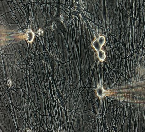
Neuroservices-Alliance is a world-renowned leader in patch clamp electrophysiology. Patch Clamp allows in vitro evaluation of compounds under the most physiologically relevant conditions.

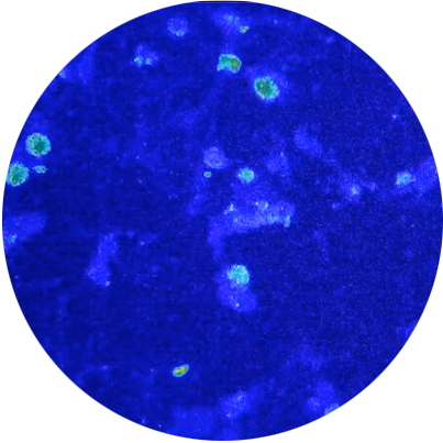
Calcium imaging provides insight into dynamic changes in calcium levels inside individual neurons. We use ratiometric calcium imaging techniques to focus on validated neurons in co-culture environments.
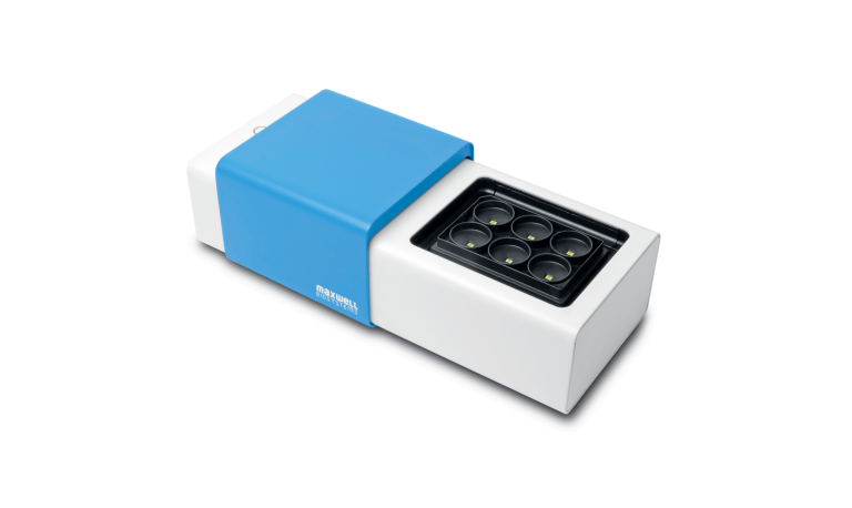
Thanks to our partnership with MaxWell Biosystems, we use the MaxTwo 6-well High Density MEA recording system. These techniques evaluate features such as global/burst firing as measures of neuronal activity across individual neurons as well as the broader network.
Download our one-pager summarizing our cell electrophysiology solutions for CNS and Pain drug discovery.
Learn more"Pharmacology of TTX-resistant and TTX-sensitive sodium currents in Non Human Primate Dordal Root Ganglia neurons."
Learn more"Profiling the Functional Phenotype of Dorsal Root Ganglia Sensory Neurons from the K/BxN Murine Rheumatoid Arthritis Model"
"Functional endpoints from human iPSC-derived sensory neurons for pain drug discovery"
Poster presented at ISSCR 2021
"High density MEA recording of primary rat neuron cultures and human iPSC-derived neuron cultures growing at low density on astrocyte feeder layers" Presented at Maxwell Biosystems Neuronal Summit in 2023
Learn more