
The VTA is a heterogeneous brain structure, containing multiple neuronal populations, including dopaminergic, GABAergic, and glutamatergic neurons. To investigate the activity of dopaminergic neurons in the VTA, stable and long-lasting recordings are performed using the MEA (multielectrode array) technique. Pharmacological isolation of dopaminergic neurons is achieved using Baclofen, a selective GABAB receptor agonist, which inhibits the firing of VTA dopaminergic neurons and a subset of glutamatergic neurons but does not affect GABAergic neurons (Margolis et al., 2012). This property allows for the selection of MEA electrodes detecting dopaminergic neuron activity.
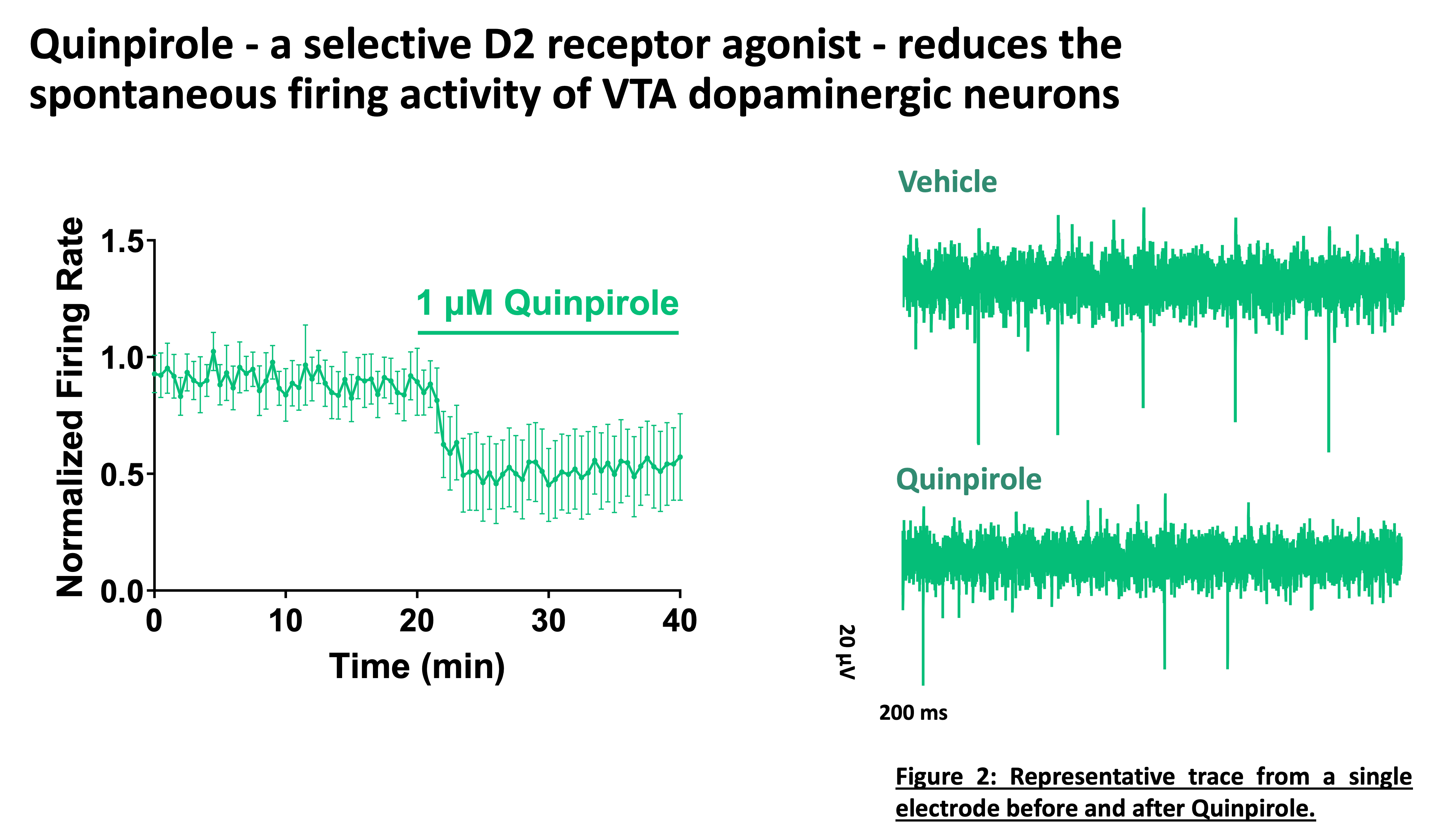
Studying the activity of these neurons provides valuable insights into the regulation of dopaminergic transmission and its modulation by pharmacological agents. Quinpirole, a D2 receptor agonist, significantly reduces the spontaneous firing of dopaminergic neurons, confirming the role of D2 autoreceptors in modulating their excitability. Additionally, the VTA receives excitatory orexinergic projections from the lateral hypothalamus, which plays a crucial role in modulating dopaminergic neuron activity. The application of Orexin-A increases the firing rate of these neurons in a dose-dependent manner, an effect specifically blocked by suvorexant, a dual orexin receptor (Ox1/Ox2) antagonist.


At Neuroservices Alliance, we provide high-quality electrophysiology data to support your preclinical research. Ready to advance your studies? Contact us today to discuss how our expertise can help drive your drug development forward!
Margolis EB, Toy B, Himmels P, Morales M, Fields HL. Identification of rat ventral tegmental area GABAergic neurons. PLoS One. 2012;7(7):e42365. doi: 10.1371/journal.pone.0042365. Epub 2012 Jul 31. PMID: 22860119; PMCID: PMC3409171.
Gotter AL, Webber AL, Coleman PJ, Renger JJ, Winrow CJ. International Union of Basic and Clinical Pharmacology. LXXXVI. Orexin receptor function, nomenclature and pharmacology. Pharmacol Rev. 2012 Jul;64(3):389-420. doi: 10.1124/pr.111.005546. PMID: 22759794.
Fadel J, Deutch AY. Anatomical substrates of orexin-dopamine interactions: lateral hypothalamic projections to the ventral tegmental area. Neuroscience. 2002;111(2):379-87. doi: 10.1016/s0306-4522(02)00017-9. PMID: 11983323.