
NS USA ran an internal methods-optimization study to answer a practical question directly: how long does it actually take, and what electrophysiological signatures tell you the patch is ready?
Whole-cell patch clamp was established on rat E18 cortical neurons, then converted to gramicidin-perforated configuration (25 ug/mL gramicidin, freshly prepared every 2-3 hours). Instead of recording at a single fixed time point, each cell was tracked continuously from patch formation through 33 minutes of perforation, with GABA currents elicited by 10 uM GABA puffs (holding potential -100 mV to +20 mV) at regular intervals (0, 5, 10, 15, 20, 25, 30, 33 min). At each time point, four parameters were measured in the same cells: access resistance (Ra), membrane capacitance (Cm), resting membrane potential (RMP), and GABA current amplitude – allowing a direct, within-cell view of how the perforated-patch configuration matures over time, rather than a single-point snapshot.
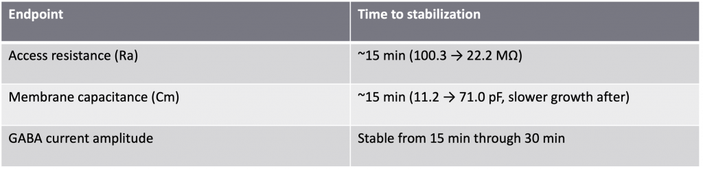
Access resistance (Ra) dropped sharply as gramicidin inserted into the membrane: from a mean of 100.3 MOhm immediately after seal formation (n=16) to 43.6 MOhm by 5 minutes, and down to 22.2 MOhm by 15 minutes (n=13). Beyond 15 minutes, Ra continued to decrease only modestly (20.0 MOhm at 20 min, 18.4 MOhm at 25 min), indicating the bulk of pore insertion is complete by the 15-minute mark.
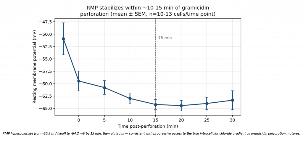
Membrane capacitance (Cm) showed the mirror-image pattern, rising as expected with progressive membrane perforation: from 11.2 pF at time 0 (n=16) to 38.8 pF at 5 minutes, then climbing more gradually to 71.0 pF at 15 minutes and 75.5 pF at 20 minutes (n=13 and n=13 respectively) – consistent with the fastest phase of pore insertion happening in the first 5 minutes, then continuing at a slower, steadier rate. Both parameters therefore converge on the same conclusion from two independent electrical signatures: by 15 minutes post-perforation, access resistance has dropped to a low, stable range and capacitance has reached a plateau-like rate of increase – the two best individual predictors of a good gramicidin-perforated recording configuration. Endpoint | Time to stabilization Access resistance (Ra) | ~15 min (100.3 -> 22.2 MOhm) Membrane capacitance (Cm) | ~15 min (11.2 -> 71.0 pF, slower growth after) GABA current amplitude | Stable from 15 min through 30 min Resting membrane potential (RMP) and GABA current amplitude were also tracked across the same time course and showed consistent stabilization once Ra and Cm reached their plateau, reinforcing that these two easily-monitored electrical parameters are reliable, real-time indicators of when a cell is ready for a trustworthy E_GABA measurement – without needing to guess or wait an arbitrary fixed time.
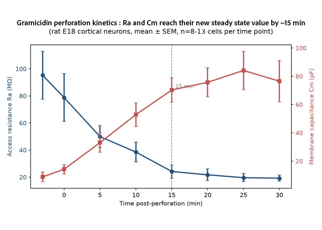
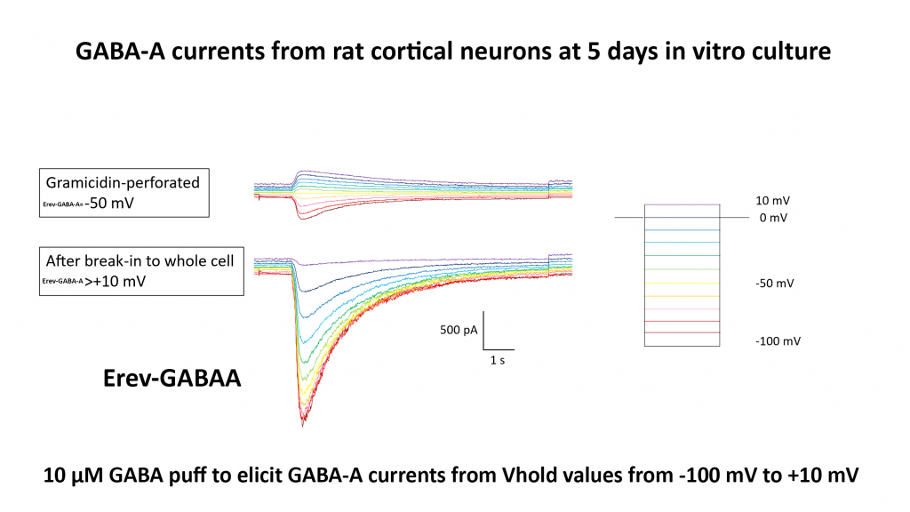
Gramicidin-perforated patch clamp for GABA current / E_GABA measurement in rat cortical neurons reaches an electrically stable, trustworthy configuration by approximately 15 minutes post-perforation, as shown convergently by access resistance and membrane capacitance kinetics tracked in the same cells over time. Rather than recording at an arbitrary fixed time point, NS USA uses Ra and Cm as real-time, per-cell quality-control criteria – waiting for access resistance to drop into a low, stable range and capacitance to plateau before running the E_GABA current-voltage curve. This directly addresses the most common failure mode of this assay: an E_GABA measurement that looks ambiguous or fails to reproduce an expected pharmacological effect not because the biology isn’t there, but because the recording itself wasn’t ready yet.