Cortical microcircuits normally balance excitatory and inhibitory (GABAergic) synaptic drive to keep network activity within a functional range. A neuromodulator that “decreases excitability” could, in principle, act through several distinct mechanisms: by directly hyperpolarizing the neuron, by reducing its intrinsic firing machinery, or indirectly, by boosting the GABAergic inhibitory tone already present in the network. Distinguishing between these possibilities matters for drug discovery — a compound that only works by amplifying existing GABAergic inhibition will behave very differently in a diseased or altered inhibitory circuit than one with a direct, GABA-independent action on the neuron.
Somatostatin receptors are expressed in cortical circuits, and somatostatin’s inhibitory effect on neuronal firing is well established pharmacologically. What is less trivial to establish in a given assay is whether blocking GABA-A receptors — the main fast inhibitory synaptic receptor in the culture — abolishes the effect (meaning it depends on the local GABAergic network) or leaves it intact (meaning the action is direct on the recorded neuron, independent of that circuit).
Using whole-cell current-clamp recording (the electrophysiology technique that tracks a neuron’s resting membrane potential, Vm, and its spontaneous action potential firing in real time), we asked:
1. Does the vehicle (carrier solution) alone have any effect on resting Vm or spontaneous firing? (negative control)
2. Does somatostatin reduce spontaneous action potential (sAP) firing in a concentration-dependent manner?
3. Does the somatostatin effect persist when GABA_A receptor-mediated inhibition is blocked with gabazine?
Cell model: primary cortical neurons dissociated from embryonic rat (E18), cultured in vitro.
Technique: Whole-cell patch-clamp electrophysiology in current-clamp mode was used to directly record membrane potential and spontaneous action-potential firing.
Protocol:Sequential drug application across multiple recording periods, referred to here as liquid periods (LPs). Four parallel experimental arms:
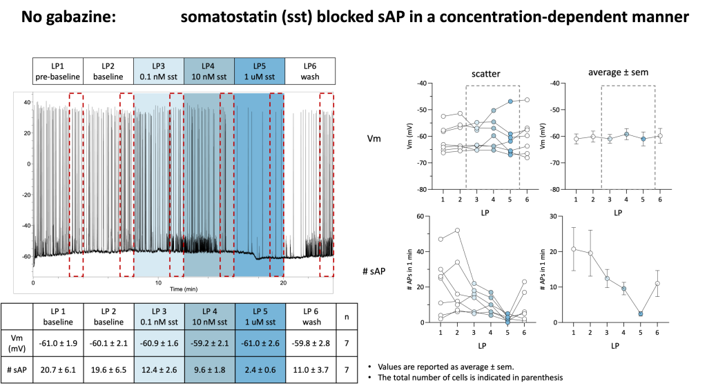
• Somatostatin, no gabazine: baseline → baseline → somatostatin 0.1 nM → 10 nM → 1 µM → wash (n = 7).
• Vehicle, no gabazine: baseline → baseline → vehicle → vehicle → vehicle → wash (n = 6).
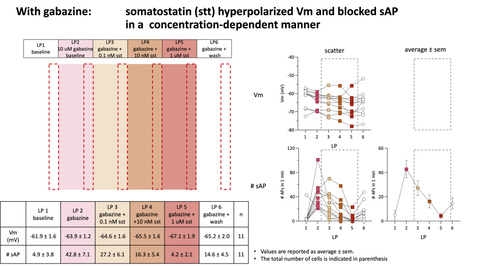
• Somatostatin, with gabazine: baseline → gabazine (10 µM) → gabazine + somatostatin 0.1 nM → 10 nM → 1 µM → gabazine + wash (n = 11).
• Vehicle, with gabazine: baseline → gabazine → gabazine + vehicle (×3) → gabazine + wash (n = 12).
Readouts: Number of spontaneous action potentials recorded during each defined analysis window.
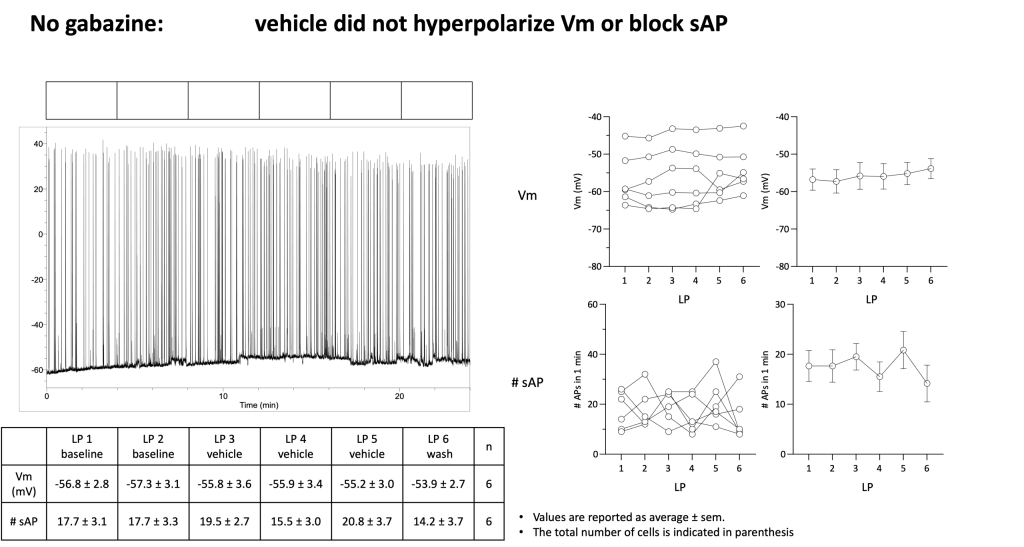
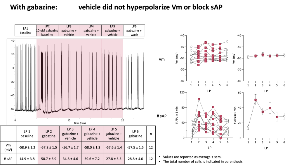
Vehicle controls, with or without gabazine, showed no apparent effect on resting membrane potential or spontaneous firing, and both measures remained stable over the recording period.
Somatostatin, no gabazine: concentration-dependent reduction of sAP firing (from 20.7 ± 6.1 APs at baseline down to 2.4 ± 0.6 APs at the highest concentration tested, 1 µM), without a significant change in resting Vm.
Somatostatin, with gabazine: concentration-dependent reduction of sAP firing was preserved (42.8 ± 7.1 APs under gabazine alone, down to 4.2 ± 2.1 APs with somatostatin 1 µM + gabazine) — and under this condition, a concentration-dependent hyperpolarization of Vm was also observed (from −63.9 ± 1.2 mV to −67.1 ± 1.9 mV).
Concentration-response fitting: somatostatin’s inhibition of sAP firing was well described by a sigmoidal fit both without gabazine (IC50 = 61 nM, Hill slope 0.59) and with gabazine (IC50 = 34 nM, Hill slope 0.64). The Vm hyperpolarization observed under gabazine also fit a sigmoidal curve (IC50 = 13 nM, Hill slope 0.47); no equivalent Vm fit could be derived in the absence of gabazine, since Vm did not change significantly in that condition.
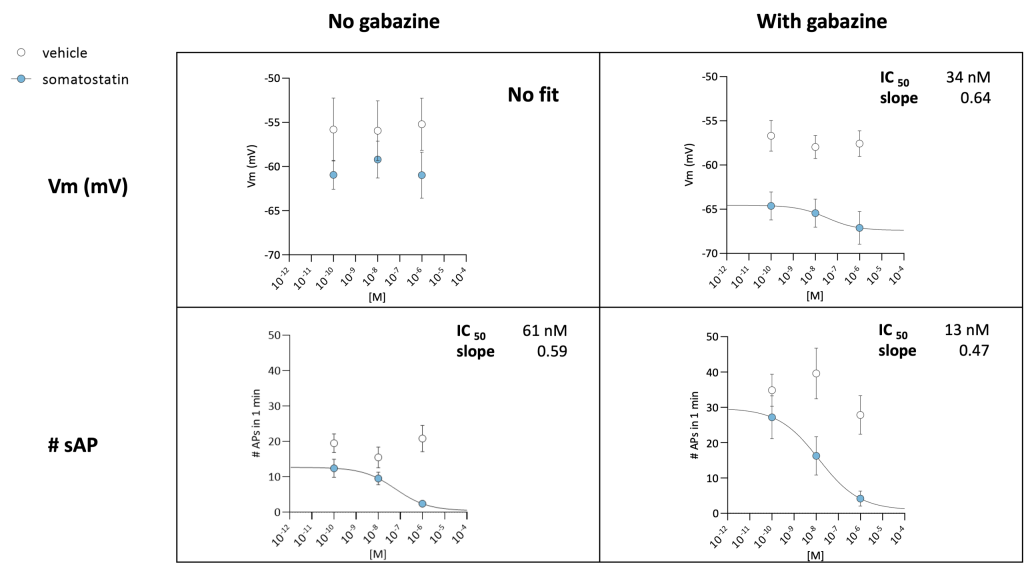
Somatostatin decreases spontaneous firing in rat cortical neurons in a clear, concentration-dependent manner — and critically, this suppression of excitability persists when GABA-A receptor-mediated synaptic inhibition is blocked. Somatostatin’s inhibitory effect persisted during GABA_A receptor blockade, supporting a GABA_A-independent effect on neuronal firing.