
The experiment recorded miniature excitatory postsynaptic currents, or mEPSCs — spontaneous electrical events that occur when a single vesicle (“quantum”) of neurotransmitter is released from a presynaptic terminal, triggering a discrete current in the recipient neuron. Because these events occur independently of action potential firing, they provide a valuable metric for assessing baseline, spontaneous synaptic transmission.
Two specific biophysical properties of mEPSCs were compared between the untreated (vehicle) control and BDNF-treated groups:
To investigate the effects of BDNF on spontaneous synaptic transmission, we recorded mEPSCs on E18 primary neurons. BDNF treatment significantly increased mEPSC frequency compared to the vehicle control group (p = 0.037; 95% CI: 0.06 to 1.67 Hz; R² = 0.122; unpaired t-test). In contrast, mEPSC amplitude showed no statistical difference between the groups (p = 0.397; 95% CI: −2.39 to 5.88 pA; R² = 0.021; unpaired t-test). The findings that BDNF increases mEPSC frequency without changing amplitude is consistent with increased spontaneous glutamate release and/or a greater number of functional excitatory synapses. This pattern, however, does not, by itself, demonstrate a purely presynaptic site of action.
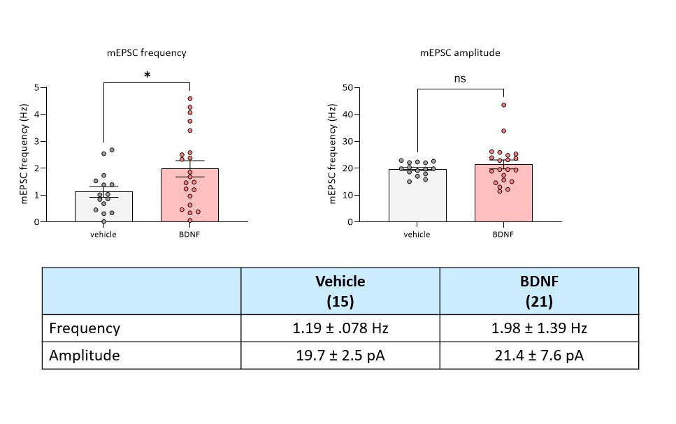
A selective change in mEPSC frequency without a corresponding shift in amplitude strongly implicates a presynaptic modification—such as an increased rate of spontaneous synaptic-vesicle fusion, elevated vesicle release probability, or a higher density of active functional release sites. Conversely, changes in mEPSC amplitude typically indicate postsynaptic adaptations, such as an alteration in the density, conductance, or sensitivity of AMPA receptors clustered at the postsynaptic density.
These biophysical readouts allow researchers to map neuromodulation on either side of the synaptic cleft. Here, BDNF’s specific modulation of frequency aligns cleanly with its established role in facilitating presynaptic neurotransmitter release and driving synaptic plasticity.
High baseline variability is fundamentally common in mEPSCs recordings from primary cultures. As is standard in preclinical electrophysiology, the necessary step requires hypothesis testing — such as an independent-samples t-test — alongside further experimental replication across additional cells and independent cultures to validate the effect.
In this dataset, BDNF treatment was associated with a higher frequency of miniature excitatory synaptic events, whereas no statistically significant difference in their mean amplitude was detected. The result provides an initial functional indication of altered spontaneous excitatory transmission, compatible with increased presynaptic release and/or increased functional synapse number.
Complementary measurements—such as paired-pulse ratio, failure rate, coefficient-of-variation analysis, synaptic-vesicle imaging or synapse-density quantification—would be required to establish the underlying mechanism(s).
In summary, the biological observation is credible and literature-compatible, including reports of BDNF potentiation in rat visual cortex and hippocampal preparations, but the document should replace “demonstrates/localizes a presynaptic mechanism” with “is consistent with a presynaptic and/or synapse-number effect.”