
Retigabine is a Kv7 (KCNQ) potassium channel opener, originally developed as an anticonvulsant. Kv7 channels carry what’s known as the “M-current,” a slow potassium current that acts as a brake on neuronal excitability, it keeps the resting membrane potential more negative and makes it harder for a neuron to fire repetitively once triggered. By opening these channels, retigabine is expected to hyperpolarize neurons and raise the threshold needed to trigger firing, effectively turning down a neuron’s overall excitability. This experiment tested that expectation directly in human iPSC-derived sensory neurons, using a protocol specifically designed to isolate multiple aspects of intrinsic excitability in the same cell.
The experiment used a rheobase protocol, run after establishing a whole-cell current-clamp recording. In this protocol, a series of 1-second depolarizing current injections of increasing amplitude is delivered to each neuron, which yields three distinct measurements from the same cell:
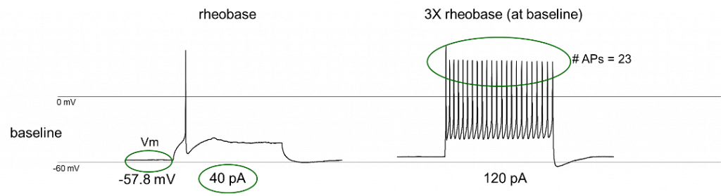
Each cell was recorded across a baseline period, a drug (or vehicle) test period, and a wash period, so that any change could be attributed to the drug and checked for reversibility. This was repeated across a concentration series of retigabine (0.3, 1, 3, 10, and 30 µM) alongside a vehicle control, allowing both a qualitative look at representative traces and a full concentration-response analysis across all three endpoints.
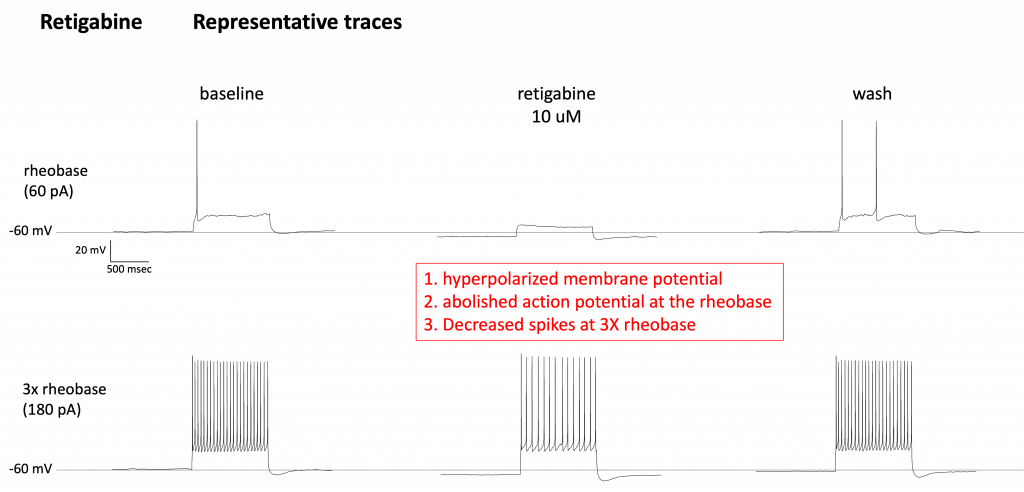
The representative traces make retigabine’s effect visually clear. At baseline, a 60 pA current injection (this cell’s rheobase) reliably triggers a single action potential, and a 3x rheobase injection (180 pA) drives a dense train of spikes. With 10 µM retigabine applied, the same 60 pA injection no longer triggers any action potential at all. The membrane potential has become more hyperpolarized, effectively raising the threshold needed to fire. At 3x rheobase, the spike train is visibly thinned out compared to baseline. After washout, firing at the rheobase current partially returns, indicating the effect is reversible rather than reflecting cell damage. Vehicle-treated cells, run through the same baseline-test-wash design, show no meaningful change in membrane potential, rheobase, or spike count at any point, confirming that the retigabine traces reflect a genuine drug effect rather than an artifact of the recording paradigm itself.

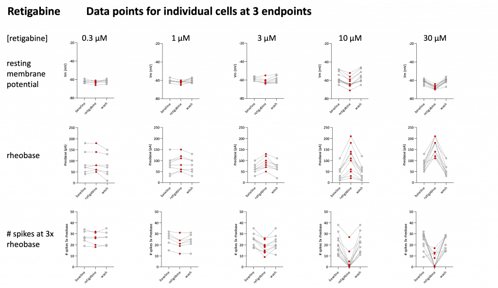
Looking at individual cells across the full concentration range reinforces this pattern and shows it scaling with dose. At low concentrations (0.3–1 µM), resting membrane potential, rheobase, and spike counts stay close to baseline values across most cells. At higher concentrations (10–30 µM), the shift becomes much more consistent and pronounced across cells: membrane potentials become more negative, rheobase increases substantially (meaning more current is needed to trigger a spike), and the number of spikes fired at 3x rheobase drops.
Fitting this data to concentration-response curves for each endpoint gives a consistent, quantified picture of retigabine’s potency:
| Endpoint | Emax | EC50 |
|---|---|---|
| Resting membrane potential | −7.7 mV | 4.8 µM |
| Rheobase | +98 pA | 7.0 µM |
| Spikes at 3x rheobase | −19 spikes | 4.2 µM |
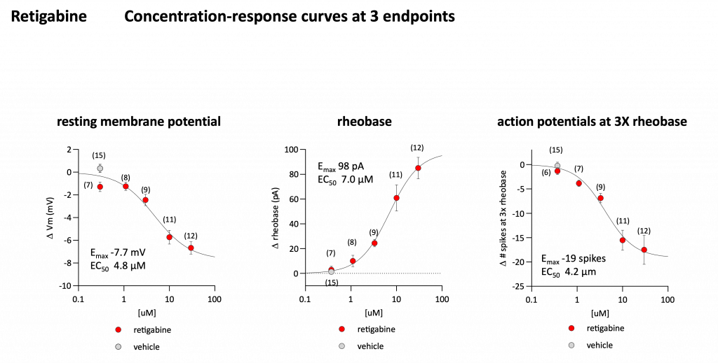
All three endpoints converge on a similar potency range (EC50 roughly 4–7 µM), which is a reassuring internal consistency check: a single mechanism — Kv7 channel opening — driving membrane hyperpolarization, raising the current threshold to fire, and reducing sustained firing, all in a coordinated, concentration-dependent way.
Across representative traces and full concentration-response analysis, retigabine consistently reduced the intrinsic excitability of human iPSC-derived sensory neurons, hyperpolarizing resting membrane potential, raising the rheobase, and decreasing the number of action potentials fired under sustained drive, all with EC50 values clustered between roughly 4 and 7 µM. That convergence across three independent, mechanistically distinct endpoints makes for a strong, well-supported picture of what a Kv7 opener does to a sensory neuron’s excitability at the single-cell level, and it’s exactly the kind of multi-endpoint characterization that helps de-risk a compound’s mechanism before it moves further down the pipeline.