In preclinical CNS and pain drug discovery, platform choice is governed by biology resolution rather than sheer throughput. Three primary modalities offer distinct windows into cellular and network functions: patch clamp electrophysiology, high-density multielectrode arrays (MEA), and Fura-2 calcium imaging. Patch clamp resolves individual ionic currents, supports IC50 determination, enables subunit dissection, and profiles single-cell excitability.
High-density multielectrode arrays (MEA) capture spontaneous and evoked population activity across hundreds to thousands of electrodes simultaneously, making it ideal for network-level pharmacology and larger compound sets.
Fura-2 calcium imaging tracks intracellular calcium flux across hundreds of individually resolved neurons, directly linking receptor engagement to downstream signaling.
As the NIH Assay Guidance Manual emphasizes, choosing a platform requires navigating an explicit trade-off: conventional electrophysiology limits throughput, while non-electrophysiological assays sacrifice voltage control and kinetic detail. To match the platform to your scientific question, evaluate your program against four core variables: target validation, screening, mechanism dissection, or translational confirmation, alongside specific cell model (primary cortical neurons, DRG sensory neurons for nociception, and human iPSC-derived neurons).
Three recording approaches dominate cell electrophysiology in preclinical CNS and pain programs, and each answers a distinct class of biological questions.
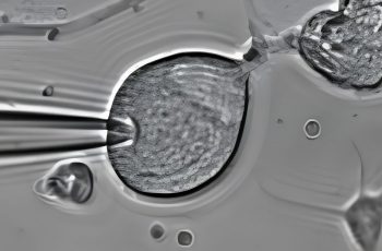
Manual patch clamp delivers the highest resolution. By establishing direct voltage control over a single neuron or transfected cell, it resolves individual ionic currents, supports precise IC50 determination, enables subunit dissection of a channel complex, and profiles single-cell excitability parameters like firing threshold, rheobase, adaptation. The NCBI Assay Guidance Manual makes the tradeoff explicit: traditional electrophysiology is low throughput, yet non-electrophysiological surrogates give up voltage control and the kinetic information that comes with it.
High-density multi-electrode array (MEA) shifts the focus from the single cell to the circuit. By recording spontaneous or evoked population activity across hundreds to thousands of electrodes at once, MEA captures firing rate, burst structure, and network synchrony. These network-level phenotypes are critical for assessing seizure liability, excitability shifts, and screening cascades where throughput is the constraint.
Fura-2 calcium imaging bridges the gap between the two. Ratiometric loading reports intracellular calcium across hundreds of individually identified neurons within a single field of view. The method excels at agonist and antagonist profiling receptors and sensory channels — such as TRP family targets in DRG neurons and allows responder-fraction analysis that single-cell recording cannot efficiently achieve.
Ultimately, these platforms are complementary, not substitutes. A hit that shifts an MEA burst pattern still requires patch clamp to identify the underlying conductance responsible. Conversely, a clean IC50 on a cloned channel says little about how a compound reshapes network firing. Robust study designs usually pair at least two of these modalities— extracting precise mechanisms from one, and population context from another.
Automated patch clamp emerged to solve a specific bottleneck in drug discovery. As the NCBI Assay Guidance Manual explains, conventional recording is inherently low throughput, while the non-electrophysiological alternatives — such as binding assays, ion flux assays, membrane-potential dyes — give up voltage control and the kinetic detail that defines behavior. Plate-based instruments were built to recover some of that critical information at a compound-screening scale. IA 2024 review of automated patch clamp places Automated patch clamp technology squarely within the realms of hit screening, target validation, and cardiac safety testing, with possible extension toward high throughput organellar recordings and optogenetic stimulation. That is the right framework: automation earns its place when the question is throughput, run-to-run reproducibility, or the early ranking of extensive many compounds against a recombinantly expressed target.
Yet, it does not answer every question. Manual recordings remain necessary when experimental protocols must be dynamically adapted at the rig — such as pre-pulse families, tail-current analysis, and use-dependence at physiological firing rates. Manual intervention is also vital when working with difficult, heterogeneous preparation like dissociated DRG sensory neurons or human iPSC-derived neurons, and whenever channel gating, kinetics or synaptic events requires expert interpretation rather than a single concentration–response metric.
Recognizing this divide, Neuroservices-Alliance delivers manual patch clamp primarily, by PhD-level electrophysiologists, choosing to absorb screening scale through high-density multielectrode array (MEA) recording and Fura-2 calcium imaging instead. In cell electrophysiology, throughput and mechanistic resolution are separate purchases; a strong study design must dictate which one you are buying.
The platform question is inseparable from the model question. Primary rat cortical neurons (E18) remain the CNS reference culture because they are synaptically active and well characterized across voltage-clamp, current-clamp, and network recordings — which makes them the practical choice for synaptic transmission, network maturation, and plasticity endpoints. DRG sensory neurons sit at the other end of the design space: they are the working model for pain pharmacology and excitability readouts, and species selection carries real pharmacological weight. Neuroservices-Alliance reports that cynomolgus monkey and dog DRG neurons give markedly different responses to Nav1.8 blockers despite similar biophysical properties — a critical translational nuance that would be entirely invisible in an oversimplified recombinant line. Human iPSC-derived neurons are the model to reach for when human biology is the deciding variable, including gene therapy validation and maturation studies spanning roughly weeks 2 to 8 in vitro.
Readout follows from mechanism, not habit. Isolating a single ionic current, dissecting subunit contributions, or generating an accurate IC50 requires the precise voltage control of manual patch clamp; the NCBI Assay Guidance Manual notes that non-electrophysiological assays cannot supply that control or the associated kinetic information. A spontaneous population firing across hundreds to thousands of electrodes is an HD-MEA question. TRP channel and G protein-coupled receptor pharmacology in sensory neurons often read out best through Fura-2 calcium imaging across many identified cells per field.
Protocol context can move the answer: in mixed cultures, blocking GABAergic tone with gabazine sharpened both the potency and efficacy of somatostatin by stripping the network’s natural inhibitory brakes. Inheriting another program’s assay design without adjusting for these microcircuits dynamics is how crucial pharmacological effects get missed.
A practical sequence uses each platform where it carries the most information. Start broad: high-density microelectrode array (MEA) recordings of spontaneous population activity across hundreds to thousands of electrodes, or Fura-2 calcium imaging across hundreds of individually identified neurons per field of view, will flag active compounds and rank them quickly. Calcium imaging is especially well suited to TRP channel and GPCR pharmacology in sensory neurons. Then, confirm mechanism at single-cell resolution with patch clamp — isolating individual ionic currents for IC₅₀ determination, subunit dissection, and excitability profiling under voltage control. The NCBI Assay Guidance Manual makes the reason explicit: non-electrophysiological readouts lack voltage control and the kinetic detail that defines how a compound acts.
That ordering is what separates initial target engagement from downstream consequence. A shift in network burst rate may reflect direct channel block, altered synaptic plasticity, or a change in inhibitory tone. Published assay notes from Neuroservices-Alliance’s report that compounds like somatostatin can produce a minimal inhibitory effect in mixed cultures but gain better efficacy and potency once background GABAergic tone is masked with gabazine — the same molecule yielding different readout depending on the circuit architecture around it. One readout rarely resolves this; complex excitability and calcium dynamics always require strategic cross-checking.
The tradeoff is real. Three complementary recording platforms demand tighter study design, and manual patch clamp trades throughput for resolution. Combined cell electrophysiology workflows repay that cost only with PhD-level scientific oversight designing them.
What to ask vendors before selecting a cell electrophysiology Partner
Instrument inventories are easy to publish; true assay validity is hard to prove. The questions below separate from a vendor that owns a rig from a team capable of answering your biological question.
Which platform is validated for my specific readout and model? Ask for the endpoint, not the instrument: single-channel currents and IC50 determination in a heterologous system are a completely different problem from spontaneous network activity in primary cortical cultures, or capsaicin-evoked calcium transients in DRG sensory neurons. The NCBI Assay Guidance Manual makes the tradeoff explicit — non-electrophysiological assays gain throughput but give up voltage control and fine kinetic detail.
Can one team run more than one platform in a coordinated design? Patch clamp, multielectrode array (MEA) recording, and fluorescence imaging confirm each other rather than substitute for each other. Ask exactly how a hit flagged on an HD-MEA system gets followed up at single-cell resolution, and who writes the final translational reports.
What is the protocol context and who signs off on the data? Request the actual protocol, the acceptance criteria, the internal control compound data, and the specific training of the scientist analyzing the raw electrophysiological traces.
What has this group published? In CNS and pain drug discovery, peer-reviewed output in electrophysiology is the most checkable proxy for technical competence.
Vendors built around automated patch clamp platforms are genuinely stronger on sheer well count for early hit screening and cardiac safety panels — a use case the 2024 review of automated patch clamp in drug discovery documents directly. The tradeoff is lower flexibility for difficult preparations and specialized protocol adaptation. Conversely, Neuroservices-Alliance position manual patch clamp for resolution target verification, paired with high-density MEA and Fura-2 imaging workflows designed around the native mechanism.