Neuroservices-Alliance (NsA) has made a significant breakthrough in CNS electrophysiological-based assays with the development of an innovative analysis tool using Artificial Intelligence (deep learning models).
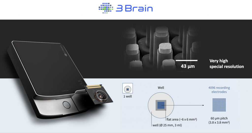
This AI algorithm offers a novel approach for spike sorting, enabling the detection of the smallest spikes with accuracy and efficiency. This tool overcomes the limitations of traditional spike sorting softwares. It provides researchers with an automated analyzing tool for data recorded with High-Density Multi-Electrode Arrays (HD-MEAs) provided by 3Brain, a Swiss-based company specializing in advanced technology for neuroscience research.
It is necessary to separate the electrical signals generated by individual neurons from the mixed signals that are recorded by electrodes because when multiple neurons are present around a same electrode, their electrical signals can overlap and become difficult to distinguish.
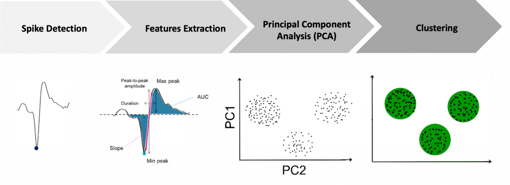
Spike sorting typically involves three main steps: spike detection, feature extraction, and clustering.
One of the main challenges in spike sorting is dealing with the variability of spike waveforms, which can be affected by factors such as electrode placement and the firing rate of neurons. Another challenge is managing the large amount of data that can be generated by high-density multi-electrode arrays, which can have hundreds or even thousands of channels.
Analyzing data from HD-MEAs can be a daunting task due to the massive amount of data generated in a short time. In a single hour, the recording from 4,096 electrodes can produce up to 1,000 GB of raw data. This raises significant challenges in terms of data management, processing, and analysis, which require sophisticated computational tools.
To overcome this challenge, NsA used a deep learning-based analysis tool to sort the spikes generated by the neurons. This tool uses 1D convolutional neural networks (CNNs) to accurately detect and sort the spikes, allowing for efficient analysis of the large amount of data generated by the HD-MEA system.
The use of deep learning models also requires significant computational power, which was achieved through the use of high-performance graphics processing units (GPUs). This combination of innovative analysis tools and powerful computing resources enables the rapid and efficient analysis of the large amounts of data generated by the HD-MEA system.
To demonstrate the effectiveness of this system, NsA conducted a study to document the effects of baclofen in cerebellum slices of 4-5 week-old mice, utilizing the AI-based solution for analysis. The AI-based analysis revealed pharmacological differences in the response of neurons to baclofen according to their location in lobules of the cerebellum.
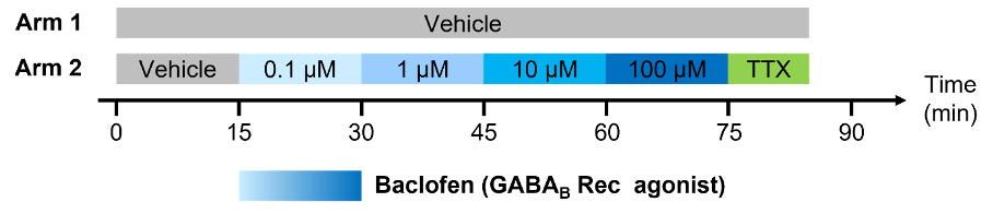
Effects of baclofen in cerebellum slices of 4-5 week-old mice
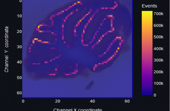
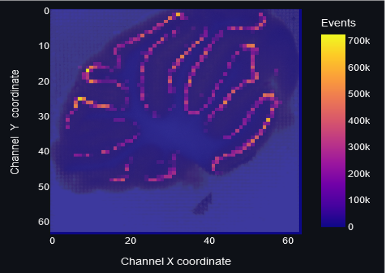
The results of the experiment using the AI-based solution revealed an impressive map of spikes detected over the course of 80 minutes, with a total of 65 million spikes detected across 417 electrodes. These spikes were sorted into 997 distinct units, providing a detailed and comprehensive understanding of the neural activity occurring during the experiment. To illustrate the analysis work, the map was color-coded to represent the number of detected events. These results highlight the power and effectiveness of the AI-based solution developed by NsA in detecting and analyzing neural activity in acute brain slices
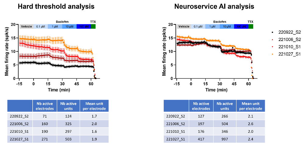
The comparison between the AI-based solution and classical methods showed that the NsA AI-based solution was superior in detecting small spikes and generating more homogeneous and robust data. This revealed that the automated analysis was very consistent and very reliable. Importantly, both methods showed the same pharmacological results, suggesting that the AI-based solution can provide the same level of accuracy as classical methods while offering additional benefits such as greater sensitivity and more homogeneous data that is independent of the operator independent.
In addition, the NsA AI-based solution demonstrated superiority in detecting more active electrodes and units compared to the classical “manual” methods. This increased sensitivity of the AI-based solution is crucial step towards a more comprehensive analysis of neural activity and the identification of subtle pharmacological properties of compounds. This could be particularly significant for the understanding of the mechanisms of action of new CNS drugs under development.
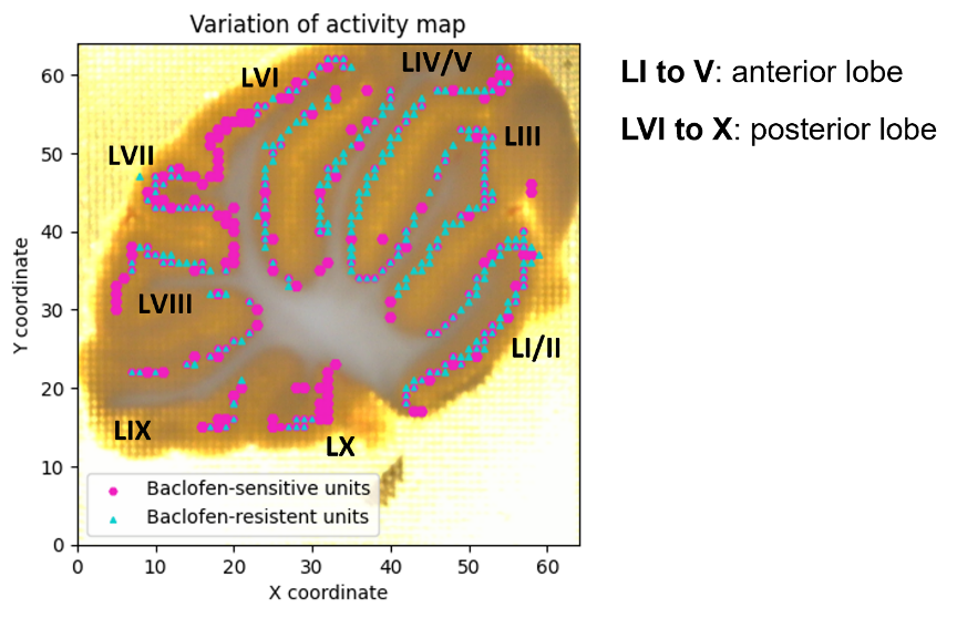
NsA’s AI-based solution, in combination with HD-MEA technology, allowed for a detailed anatomical analysis of a drug effect in cerebellum, a region of the brain responsible for motor control, coordination and decision making. The AI-based analysis revealed that the posterior lobe of the cerebellum is more sensitive to baclofen. In contrast, the anterior lobe of the cerebellum was found to be a little less sensitive to the drug.
The ability to identify such subtle differences in drug sensitivity across different regions of the brain can have important implications for the development of targeted therapies for neurological disorders. By understanding which regions of the brain are most affected by a particular drug, researchers can develop more effective treatment strategies that minimize side effects and maximize therapeutic benefits.
The AI-based solution also offers several other advantages for researchers. One of the key benefits is the ability to group units based on their anatomical location, allowing for fine anatomical analysis with strong statistical power and subregional analysis.
Furthermore, the automated nature of the analysis with the AI-based solution reduces the potential for human error and increases the speed and efficiency of data analysis. This allows researchers to focus on the interpretation of the results, rather than spending significant time on manual data analysis.
Custom-made solutions developed by NsA provide flexibility and on-demand analysis and graphical representations, allowing NsA researchers to tailor their analysis to specific needs and extract the maximal information for its customers compound.

Saber Graf
Software Developer

Benoît Buisson
Software Developer

Julien Artinian
Electrophysiology Engineer
Are you interested in exploring our AI analysis tools for HD-MEA recordings and discover how our custom-designed protocols could be of interest for your ongoing CNS Research?